All biopsy and fluid samples are sent to a pathologist. Pathologists use microscopes and special stains and assays to find out if the samples contain mesothelioma.
Microscope not sufficient
Under the microscope, mesothelioma may look like other types of cancer. For example, pleural mesothelioma can resemble some types of lung cancer, and peritoneal mesothelioma in women may look like some types of ovarian cancer. Because of this, special tests and assays are used by pathologists. These tests actually use special stains called immunohistochemistry to look for tell-tale proteins (antigens) often over expressed in mesothelioma cells.
Immunohistochemical Assay for Pleural Mesothelioma
For example, here are some immunohistochemical markers used to help identify epithelioid pleural mesothelioma:
- Calretinin – Demonstrated in virtually all epithelioid mesotheliomas. The staining is often strong and diffuse.
- Keratin 5/6 – It is expressed in 75%–100% of the mesotheliomas.
- WT-1 protein – Forty-three percent to 93% of the mesotheliomas show nuclear positivity.
- Podoplanin (D2-40) – Eighty-six percent to 100% of mesotheliomas show positivity along the cell membranes.
Immunohistochemical Assay for Peritoneal Mesothelioma
Here are some immunohistochemical markers used to help identify peritoneal mesothelioma:
- Calretinin – Positive in 85%–100% of peritoneal mesothelioma cases.
- D2-40 – Positive in 93%–96% of peritoneal mesothelioma cases.
Very often, additional markers are used to test for other cancers. So for example, the pathologist will run an assay that uses specific markers that are sensitive to mesothelioma cells and other markers that are specific to other cancers. Based on the results, the pathologist can distinguish one cancer from another. That is why if you look at your pathology report, very often it will show positivity for some tests and negativity for others.
Mesothelioma Cell Types
During this process, the pathologist may also determine the mesothelioma cell type. Most mesotheliomas are classified as either epithelioid, sarcomatoid, or mixed/biphasic.
Each of the different cell types not only stain differently with immunohistochemistry tests, but also look different under the microscope.
According to pathologists:
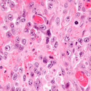
- “Epithelioid mesothelioma are composed of polygonal, oval, or cuboidal cells that often mimic nonneoplastic reactive mesothelial cells.”
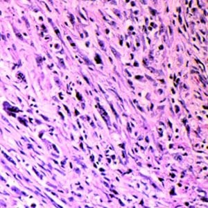
- “Sarcomatoid mesotheliomas usually consist of spindle cells but can be composed of lymphohistiocytoid cells and/or may also contain heterologous rhabdomyosarcomatous, osteosarcomatous, or chondrosarcomatous elements.”
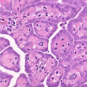
- “Mixed or biphasic mesotheliomas contain both epithelioid and sarcomatoid areas within the same tumor.”
Read more about mesothelioma cell types.
Prognosis & Survival
Epithelioid generally has the best prognosis, sarcomatoid, the worse and biphasic in the middle (because it can possess both of the other cell types).
However, it’s important to note that the prognosis associated with each cell type comes from the results of standard treatments such as chemotherapy and surgery. There is limited data on which cell type has a better prognosis when the patient uses clinical trials, and virtually no data relating cell type to prognosis when the patient uses alternative modalities.
What Next?
It is a good idea to learn about your cancer by reading your pathology report and discussing it with your doctor. For example, if you have the sarcomatoid type of mesothelioma and your doctor is recommending chemotherapy treatment, you may want to ask what the prognosis is with this type of treatment to help weigh the pros and cons.