Imaging scans are an important tool in helping clinicians diagnose and manage mesothelioma. These scans include:
- X-rays
- CT scans
- MRIs
- PET scans
These tools help doctors to actually see tumors and other abnormal growths inside a patient’s body.
When patients experience symptoms associated with mesothelioma, doctors often order a scan as one part of their work-up. The results of the scan can help doctors figure out what is causing the symptoms. A scan by itself cannot diagnose mesothelioma, but it is an important tool towards arriving at the correct diagnosis.
X-rays for Mesothelioma
X-ray is the oldest and most basic type of imaging. X-ray’s produce a flat, two-dimensional image so its capacity as a helpful diagnostic tool is limited. Nevertheless, some physicians use it as a start. If an abnormality is seen, such as a mesothelioma tumor, more accurate and sophisticated scans can then be ordered like those described below.
X-rays work be emitting electromagnetic radiation. The radiation is focused on a particular body part and a photographic film is placed on the opposite side. The rays pass through less dense tissue more easily and create an image that is dependent on what the rays encountered while traveling through the body. For example, standard X-rays portray healthy lungs as black. However, when a tumor is present on or near the lungs (such as on the pleural lining), doctors may see an opaque white area.
CT Scans for Mesothelioma
We have all heard of CT or CAT scans. These are actually computed tomography scans which is a type of X-ray. In fact, CT scans are just a more sophisticated use of X-ray technology. CT’s take many X-rays from different planes or cross-sections. Because there are so many “slices” taken at about the same time, these images can be reconstructed by a computer to create a three dimensional representation of what is inside the body. As a result, CT’s are a very useful tool when doctors are trying to see if there are any masses or tumors present such as mesothelioma, and the location and size of any abnormal growths.
Contrast Material
Often, contrast material is used to help make a CT scan as accurate as possible. Contrast materials may also be called contrast agents or contrast media and are used to improve pictures of the inside of the body. Contrast materials are basically dyes that temporarily discolor internal organs and change the way x-rays or other imaging tools interact with the body.
By improving the visibility of specific organs or tissues, contrast materials help physicians diagnose mesothelioma and other medical conditions. Contrast materials enter the body by being swallowed, injected into a blood vessel, or administered by enema. There are several types of contrast materials including iodine-based and barium-sulfate compounds. When these contrast materials are present in a specific area of the body, they block or limit the ability of x-rays to pass through. As a result, blood vessels, organs and other tissue that contain the dyes change their appearance on x-ray or CT images.
Many doctors believe that CT’s are the best imaging technology for scans of the chest and abdomen, the most common location of mesothelioma tumors. In addition to finding potential tumors, CT scans can also help doctors perform staging by determining if cancer has spread to lymph nodes, nearby tissues, or distant organs.
MRI Scans for Mesothelioma
Unlike X-rays and CT’s, magnetic resonance imaging (MRI) scans use a completely different type of radiation to develop images of the inside of the body to see mesothelioma and other cancers.
MRI’s work on an atomic scale by temporarily realigning the protons in the water molecules in your body. An MRI applies a very strong magnetic field (about a thousand times the strength of a typical refrigerator magnet), to change the spin of protons. The scanner also produces a radio frequency that creates a varying magnetic field. The protons absorb the energy from the field. When the field is turned off, the protons gradually return to their normal spin. The return process produces a signal that can be made into an image.
What’s amazing is that the protons in different body tissues return to their normal spins at different rates, so the scanner can distinguish among tissues including tumors. In fact, the computer can differentiate between tissues in the body and assign them various colors. Doctors get a clear image of the interior of the body, which can help locate mesothelioma earlier than with X-rays and CT scans.
PET Scan
A positron emission tomography (PET) scan is another technology that allows your doctor to check for diseases in your body like mesothelioma. The scan uses a special kind of dye that has radioactive tracers. Your organs and tissues absorb the tracer and when highlighted under a PET scanner, the tracers help your doctor see how well your organs and tissues are working. The PET scan can measure blood flow, oxygen use, glucose metabolism (how your body uses sugar), and much more. Because cancer cells often have a higher metabolism than normal cells, a PET scan can be useful in detecting mesothelioma and other tumors.
Unlike other imaging tests, such as CT or MRI, PET scans show problems at the cellular level. This allows your doctor to see how the cancer metabolizes, and whether it has spread, or metastasized, to new areas. PET scans can also show how a tumor is responding to chemotherapy.
Before the scan, the tracers may be delivered intravenously or through a solution you can drink. Your body needs time to absorb the tracers, so you will usually wait about an hour before the scan begins.
These are some of the amazing technologies that clinicians have to help diagnose mesothelioma and other cancers and diseases.
Related Posts
 Ultrasound More Accurate for Malignant Pleural Mesothelioma - Ultrasound may be a more accurate and cheaper option than CT scans for malignant pleural mesothelioma. A group of Italian researchers examined the use of ultrasound to help diagnose malignant pleural mesothelioma. The goal was to help doctors locate optimal sites for biopsies. Diagnosing Malignant Pleural Mesothelioma Mesothelioma is a rare form of cancer that develops from mesothelium cells. The mesothelium is the lining that covers many of our internal organs. Mesothelioma is caused by exposure to asbestos, a toxic substance used in industrial and consumer products. When mesothelioma develops in the lining of our lungs, it is called malignant pleural mesothelioma. It can take around 40 years for malignant pleural mesothelioma to develop after asbestos exposure. Diagnosing malignant pleural...
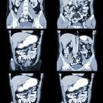 New Model for the Diagnosis of Mesothelioma - A new model studied by researchers from China could help doctors to tell two different cancers apart. Mesothelioma scan results can often be confused with another cancer, called metastatic pleural disease (MPD). Mesothelioma is a rare type of cancer that develops in the linings of internal organs. When the cancer grows in the lining of the lungs, it is called malignant pleural mesothelioma (MPM). This is because the lining the of the lungs and chest are called the pleura. MPD also forms in the lining of the lungs. This cancer starts to grow in a different organ and spreads to the lung. Unlike mesothelioma, it is not caused by exposure to asbestos. Difficulties in the Diagnosis of Mesothelioma Mesothelioma is...
 High-Resolution Imaging for Mesothelioma Staging and Diagnosis - High-resolution imaging is one of the most important tools for a clinician. A new report in the AME Surgical Journal shows the importance of better clinical imaging. It is key for accurate diagnosis and staging of malignant mesothelioma. Malignant pleural mesothelioma is a complex disease. Mesothelioma diagnosis and accurate staging are often difficult to pin down. And the diagnosis is further complicated by different disease subtypes. Higher resolution diagnostic imaging plays an important role in diagnosis and staging. It shows tumor thickness or volumetric data. Future updates to the cancer staging system may use clinical imaging benchmarks. Importance and Use of Clinical Imaging Mesothelioma causes significant morbidity and mortality. And it remains a challenging disease to diagnose and stage accurately....
 How Do We Know If the Treatment is Working? Measuring Malignant Pleural Mesothelioma Treatment Response - Malignant pleural mesothelioma is a rare type of cancer. A growing number of mesotheliomas are being treated with novel immunotherapies. But clinicians still lack a meaningful way to measure mesothelioma treatment response. An article in Seminars in Nuclear Medicine lists five ways clinicians can measure how well a treatment plan is working. And a new artificial intelligence tool can measure mesothelioma tumors without any human input. Artificial intelligence may provide the key to treatment response in the future. Measuring Mesothelioma Treatment Response In malignant pleural mesothelioma, complex tumors result in inconsistent clinical assessment. There are five common ways to measure if a treatment plan is working. Generally, a clinician will look to see if the size or weight of a tumor has...
 Clinical Scan Anxiety before a Mesothelioma Medical Scan? You’re not Alone. - Many mesothelioma patients with advanced cancer have anxiety before a clinical scan. Clinicians have even coined this clinical scan anxiety as “scanxiety.” A new study published by Supportive Care in Cancer looked at the experiences of scans and scanxiety in patients with advanced cancer. They were looking for the best strategies to reduce clinical scan anxiety or scanxiety. The research team conducted interviews with 16 patients with advanced cancer. The team asked about anxiety felt during computed tomography (CT) scans for monitoring of their cancer. Anxiety About the Scan Experience and Scan Results For these patients, scans were viewed as a routine and normal part of cancer care. This is one of those “necessary evils” that is part of the...
 Using CT Scans in Surgical Decision-Making in Peritoneal Mesothelioma - Peritoneal mesothelioma is caused by asbestos. The peritoneum is the space in your abdomen that contains the intestines, the stomach, and the liver. The abdomen (peritoneum) is the second most common site of mesothelioma after the pleural (lungs and chest). Most doctors believe peritoneal mesothelioma is caused by the ingestion of asbestos fibers. Microscopic asbestos fibers become embedded in the abdomen (peritoneum). After about 20-50 years, these fibers can cause inflammation and mutations in the healthy mesothelial cells. These mutations may ultimately cause these cells to become cancerous. Thus, forming tumors in the peritoneum. Patients diagnosed with peritoneal mesothelioma may survive several or more years after diagnosis. There are long-term survivors, such as Paul Kraus. Using CT Scans to Avoid...
 Mesothelioma and MRI Scans: Using an Old Tool in a New Way - Imaging scans are an important tool in helping clinicians diagnose mesothelioma. These scans include X-rays, CT scans, MRI scans, and PET scans. These tools help clinicians to see tumors inside a patient’s body. The results of the scan cannot diagnose mesothelioma, but it is an important tool for arriving at the correct diagnosis. Unlike X-rays and CT’s, magnetic resonance imaging (MRI) scans use a completely different type of radiation. An MRI applies a very strong magnetic field to change the spin of protons. The scanner also produces a radio frequency. This process produces a signal that can be made into an image. Until now, the role of MRIs has been limited in mesothelioma. However, new evidence suggests MRIs can be...
 New Artificial Intelligence Tool Measures Mesothelioma Tumors - A new artificial intelligence tool can measure mesothelioma tumors without any human input. Automated mesothelioma tumor measurement is now possible. The artificial intelligence algorithm performed well. There were 266 malignant pleural mesothelioma tumors analyzed. This is the largest study of its kind performed in mesothelioma. And this is the first description of an automated artificial intelligence tool. Inconsistent Mesothelioma Measurement During Clinical Assessment In malignant pleural mesothelioma, complex tumors result in inconsistent clinical assessment. Promising new methods need practical computer automation methods. A new publication showed an automated tool for this purpose. This study compared a new artificial intelligence tool with human standard clinical assessment. This study looked at mesothelioma patients treated with chemotherapy. Evaluating How Well the New Tool...
 Mesothelioma Mimics Ovarian Cancer: Lessons from a Case Report - Mesothelioma mimics ovarian cancer in a new report by Radiology Case Reports. Mesothelioma is a rare and very aggressive tumor of the peritoneum. Similar clinical and imaging presentations mean mesothelioma mimics ovarian cancer at times. Yet, a history of asbestosis exposure is key as the main risk factor for mesothelioma. Imaging plays a key role in getting the diagnosis correct. It narrows down the diagnosis possibilities. Ultrasound-guided biopsy with histological study can confirm a mesothelioma diagnosis. Case Report Misdiagnosis The Bangladesh National Institute of Oncology recently published a case report. A 42-year-old woman reported pelvic pain and progressive abdominal distension. An abdominal ultrasound revealed a high volume anechoic peritoneal effusion. Further investigation showed extensive peritoneal disease and bilateral ovarian tissue...
 Artificial Intelligence Tool Measures Mesothelioma Treatment Response - Scientists at the University of Glasgow in Scotland have developed an artificial intelligence tool that could revolutionize the way doctors plan and monitor mesothelioma treatment. The University team collaborated with Scottish firm Canon Medical Research Europe to develop an AI prototype for detecting pleural mesothelioma. Canon Medical specializes in medical imaging software. Pleural mesothelioma is a type of lung cancer caused by asbestos exposure. The new artificial intelligence tool is designed to find and measure mesothelioma tumors on CT scans. It can be challenging even for cancer experts to detect tiny changes in mesothelioma tumors. But the AI tool can quickly reveal how well or poorly a patient is doing on a treatment like chemotherapy. The sooner doctors know if...