HIPEC Treatment: Heating Up the Fight Against Mesothelioma
|

HIPEC Treatment: Heating Up the Fight Against Mesothelioma

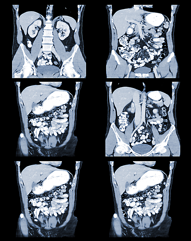
An elderly woman with malignant mesothelioma had a positive response to HIPEC treatment. HIPEC  stands for hyperthermic intraperitoneal chemotherapy. The Journey of Diagnosing Malignant Peritoneal Mesothelioma Mesothelioma is a type of cancer that is caused by exposure to asbestos. It develops in the lining of organs in the body. When it grows in the abdominal cavity, it is called malignant peritoneal mesothelioma. This type of mesothelioma causes symptoms like abdominal pain, bloating, weight loss, and fluid build-up in the abdomen. It can take decades after asbestos exposure for mesothelioma symptoms to appear, and it can be hard for doctors to diagnose this cancer. Triumph Over Malignant Peritoneal Mesothelioma with HIPEC In this case, a 64-year-old woman went to the hospital…

New Model for the Diagnosis of Mesothelioma
|

New Model for the Diagnosis of Mesothelioma

A new model studied by researchers from China could help doctors to tell two different cancers apart. Mesothelioma scan results can often be confused with another cancer, called metastatic pleural disease (MPD). Mesothelioma is a rare type of cancer that develops in the linings of internal organs. When the cancer grows in the lining of the lungs, it is called malignant pleural mesothelioma (MPM). This is because the lining the of the lungs and chest are called the pleura. MPD also forms in the lining of the lungs. This cancer starts to grow in a different organ and spreads to the lung. Unlike mesothelioma, it is not caused by exposure to asbestos. Difficulties in the Diagnosis of Mesothelioma Mesothelioma is…

Using CT Scans in Surgical Decision-Making in Peritoneal Mesothelioma
|

Using CT Scans in Surgical Decision-Making in Peritoneal Mesothelioma

Peritoneal mesothelioma is caused by asbestos. The peritoneum is the space in your abdomen that contains the intestines, the stomach, and the liver. The abdomen (peritoneum) is the second most common site of mesothelioma after the pleural (lungs and chest). Most doctors believe peritoneal mesothelioma is caused by the ingestion of asbestos fibers. Microscopic asbestos fibers become embedded in the abdomen (peritoneum). After about 20-50 years, these fibers can cause inflammation and mutations in the healthy mesothelial cells. These mutations may ultimately cause these cells to become cancerous. Thus, forming tumors in the peritoneum. Patients diagnosed with peritoneal mesothelioma may survive several or more years after diagnosis. There are long-term survivors, such as Paul Kraus. Using CT Scans to Avoid…

CT Evaluation Correlates with Actual Mesothelioma Tumor Volume, Subtype, and Survival
| |

CT Evaluation Correlates with Actual Mesothelioma Tumor Volume, Subtype, and Survival

Two new studies highlight the value of CT evaluation for patients with either peritoneal or pleural mesothelioma.  CT stands for computed tomography. The technology uses X-rays to create three dimensional images of internal structures.  The studies show CT evaluation can give doctors an accurate idea of the size and extent of mesothelioma tumors. These scans can also help predict which patients have the best odds of survival. This helps doctors plan better, more effective treatment strategies.  What is Malignant Mesothelioma? Malignant mesothelioma is a type of cancer that grows on the membranes around internal organs. The most common type of mesothelioma is pleural mesothelioma. This kind of tumor starts on the pleural membrane around the lungs.  Peritoneal mesothelioma accounts for…

Predicting Mesothelioma Outcomes with FDG-PET: A New Model
| | | |

Predicting Mesothelioma Outcomes with FDG-PET: A New Model

Swiss doctors have created a new tool for predicting mesothelioma outcomes with FDG-PET scans.  FDG-PET scanning is a nuclear imaging technique. It gives doctors a non-invasive way to track the growth of pleural mesothelioma tumors.  The Swiss team analyzed multiple PET scans and CT scans from mesothelioma patients. They used a computer to look for commonalities in the scans. Then they used the information to create a computer model for predicting mesothelioma outcomes with FDG-PET. The model may help doctors and patients make better decisions about mesothelioma treatment in the future.  Imaging Studies for Mesothelioma Prognosis Pleural mesothelioma tumors occur on the pleural lining around the lungs. In the early stages, mesothelioma causes few symptoms. As mesothelioma tumors grow, they…

Diagnostic CT Scan for Mesothelioma: More is Better
| | |

Diagnostic CT Scan for Mesothelioma: More is Better

A new report out of the UK suggests that a thorough diagnostic CT scan for pleural mesothelioma should include images of the abdomen and pelvis, as well as the chest.  When a person goes to the doctor with early signs of malignant pleural mesothelioma, a CT scan of the chest is often the next step in making a diagnosis. CT scans create a 3-dimensional image of internal organs, making it easier to spot mesothelioma tumors.  But a research team led by the University of Bristol says a diagnostic CT scan that shows only the chest might miss mesothelioma tumors in other areas. How a Diagnostic CT Scan Works CT stands for computerized tomography. CT scanning is an advanced type of…

Low-Dose CT Screening May Delay Mesothelioma Deaths
| | |

Low-Dose CT Screening May Delay Mesothelioma Deaths

The results of a long-term national study suggest that low-dose CT screening might prevent – or at least delay – deaths from pleural mesothelioma.  The National Lung Screening Trial included more than 53,000 lung cancer patients. Researchers released the first results in 2011 after more than six years of study. The recent follow-up results reflect more than 12 years of data on low-dose CT screening. They show that, in many cases, screening lung cancer patients for at least three years delayed their deaths for more than a decade.  Pleural Mesothelioma and Lung Cancer Pleural mesothelioma is not the same thing as lung cancer. Mesothelioma starts on the lining around the lungs (pleura), rather than in the lung tissue itself.  But…