CT Scans Fail To Identify Metastatic Mesothelioma Prior to Surgery

CT scans are not a reliable way to detect the spread of mesothelioma into certain lymph nodes between the ribs. This is important because people with mesothelioma cells in their posterior intercostal lymph nodes (PILN) do not tend to get good results from P/D surgery.
Surgeons need a good way to find mesothelioma cells in these lymph nodes before they decide to perform this risky operation. But University of Pennsylvania researchers say CT scans are not the best method.
CT Scans in Mesothelioma Diagnosis and Prognosis
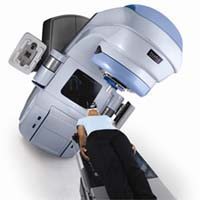
CT stands for computerized tomography. A CT machine uses a series of X-ray images from different angles to create a 3D picture of a mesothelioma tumor. Most hospitals have a CT scanner. They are more common than PET or MRI machines.
CT scans are one of the main ways that doctors identify cancer. They use a combination of imaging scans, exams, biomarker tests, and patient history to make a mesothelioma diagnosis. CT scans can also show if a treatment is working.
But CT scans have limitations, too. A CT image may show a tumor as a dark mass. But it is not always possible to tell from the picture if the mass is cancer. Doctors often combine computed tomography with PET scanning for better results.
Metastatic PILN and P/D Surgery
Pleurectomy with decortication (P/D) is one of the two main types of surgery for mesothelioma. Surgeons remove the diseased pleural membrane and other at-risk tissues.
The posterior intercostal lymph nodes are toward the back of the chest, between the ribs. Research shows that people whose mesothelioma is already in the PILN have shorter survival after P/D surgery.
The new study tests the value of CT scans for finding metastatic PILN before surgery. Thirty-six patients underwent extended P/D surgery for pleural mesothelioma between 2007 and 2013.
Comparing CT and Pathology
During surgery, doctors discovered that 22 patients had mesothelioma cells in their PILN. Fourteen patients did not. Radiologists who did not know the results of surgery analysed the preoperative CT scans.
CT scans did show some of the PILN. But most of the scans could not show if the PILN had mesothelioma cells in them.
“The number of PILN on preoperative CT did not predict metastasis with an average of 2 PILN seen, regardless of PILN pathology,” states the report in Lung Cancer.
The benign and malignant PILN were about the same size. The different radiologists found about the same number and size of PILN in the CT scans.
The report concludes, “CT does not reliably identify metastatic PILN on preoperative CT for patients with malignant pleural mesothelioma undergoing extended pleurectomy/decortication.”
Source:
Berger, I, et al, “CT for detection of malignant posterior intercostal lymph nodes in patients undergoing pre-operative staging for malignant pleural mesothelioma”, December 4, 2020, Lung Cancer, https://www.lungcancerjournal.info/article/S0169-5002(20)30720-0/fulltext


