Invasiveness of Peritoneal Mesothelioma: MRI Beats CT for Assessment
New research shows MRI beats CT when it comes to measuring the invasiveness of peritoneal mesothelioma.
Scientists from several of the top US cancer centers ran the study. It included nearly 500 mesothelioma patients and appears in the journal Clinical Colorectal Cancer.
The study shows that the invasiveness of peritoneal mesothelioma is more visible on MRI than it is on CT. The news could make a difference in how doctors plan peritoneal mesothelioma treatment.
Peritoneal Cancer Index and Mesothelioma
Peritoneal mesothelioma cancer grows on the peritoneal membrane lining the abdomen. Like all forms of mesothelioma, it usually comes from exposure to asbestos. Peritoneal mesothelioma accounts for about a fifth of all mesothelioma cases.
Doctors assess the invasiveness of peritoneal mesothelioma using the Peritoneal Cancer Index (PCI). The PCI assigns a number based on how many regions of the abdomen the cancer is in. The higher the PCI, the more invasive the cancer.
Doctors use the PCI to plan for surgery and other mesothelioma treatments. PCI can determine whether a mesothelioma patient is a candidate for CRS surgery with HIPEC.
Assessing Invasiveness of Peritoneal Mesothelioma
The only fool-proof way to determine the invasiveness of peritoneal mesothelioma is to operate. But imaging tools like MRI and CT can give doctors a pretty good idea of the PCI before surgery.
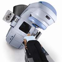
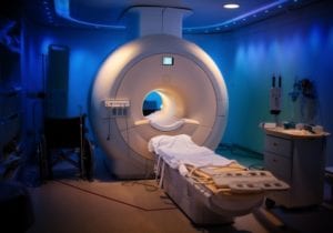
Magnetic resonance imaging (MRI) uses a strong magnet and radio waves to create an image of the inside of the body. Computerized tomography (CT) creates an image by piecing together a series of X-ray pictures.
The new study included 488 patients with peritoneal cancer. Eight percent of the patients had peritoneal mesothelioma. All study subjects underwent CT and/or MRI prior to surgery with heated intraperitoneal chemotherapy (HIPEC). The goal of the study was to see how closely the actual invasiveness of peritoneal mesothelioma matched the predictions based on imaging studies.
MRI Versus CT for Imaging Mesothelioma
Both MRI and CT images gave doctors a pretty good idea of each patient’s PCI. But some cancers seemed to be more visible with one type of imaging than the other.
For patients with appendiceal or colorectal cancer, CT imaging correlated well with the PCI. But CT was not as good at showing the invasiveness of peritoneal mesothelioma.
On the other hand, the researchers say “MRI-PCI was correlated with intraoperative-PCI for all histologies.” That includes peritoneal mesothelioma. They conclude, “MRI appears to be superior for invasive appendiceal and peritoneal mesothelioma.” A larger study will be needed to validate the idea.
The invasiveness of peritoneal mesothelioma has a bearing on surgery outcomes. The study showed that patients with a PCI above 15 tended to have less complete cytoreduction. That means doctors were not able to remove all of the cancer.
Source:
Lee, RM, et al, “What is the Optimal Preoperative Imaging Modality for Assessing Peritoneal Cancer Index? An Analysis From the United States HIPEC Collaborative”, December 12, 2019, https://www.clinical-colorectal-cancer.com/article/S1533-0028(19)30483-9/fulltext