CT Accuracy May Vary for Mesothelioma

A group of French doctors has a warning for people exposed to asbestos and concerned about the possibility of mesothelioma: watch out for false positive CT results.
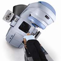
Their new meta-analysis of current radiological techniques found that there is a wide margin in the way CT results are interpreted in cases of cancer in the lungs and chest including mesothelioma. The study may have important implications for mesothelioma, where early, accurate diagnosis is critical. CT (computed tomography) and the newer high-resolution volume CT (HR-VCT) which takes multiple two-dimensional images, are among the most popular imaging modalities for diagnosing and staging malignant pleural mesothelioma.
According to the researchers who conducted the study, part of the problem is the lack of specific training in asbestos-related illnesses for radiologists, resulting in variable recognition of mesothelioma and other asbestos diseases, as well as the lack of “standardized, quantified criteria for CT abnormalities.” To ensure the accuracy of mesothelioma diagnoses, the team suggests that radiologists evaluating asbestos-exposed patients refer to a set of standardized guidelines such as the “CT Atlas of Benign Diseases Related to Asbestos Exposure”, published by a group of French experts in 2007, when making their diagnosis.
In an article in the French medical journal Review of Respiratory Maladies, the authors write, “The very low agreement between thoracic and general radiologists must be taken into account. The reading of CT scans in cases of occupational exposure to asbestos should be entrusted to thoracic radiologists or to general radiologists having validated specific training. “
Mesothelioma affects an estimated 2,000 people in the U.S. each year as well as tens of thousands of people around the world. Although it is still relatively rare, this asbestos-linked cancer is highly aggressive and often resistant to standard therapies.
Source:
Ferretti, G, “What are the tools for post-occupational follow-up, how should they be performed and what are their performance, limits and benefit/risk ratio? Chest X-Ray and CT scan” [Article in French], June 2011, Revue des Maladies Repiratoires, pp. 761-772.