Mesothelioma Staging: Laparoscopy May Find Tumors PET-CT Misses
Cancer researchers at Baylor College of Medicine say diagnostic laparoscopy (DL) improves the accuracy of pleural mesothelioma staging prior to surgery. It is especially valuable for patients with cancer in both the chest and abdomen.
Staging is a way for doctors to determine how widespread cancer is. Accurate staging is critical for the best surgical outcomes. For the right patients, mesothelioma surgery can be a life saver.
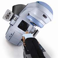
Many mesothelioma patients undergo PET/CT as part of the staging process. But the Baylor study suggests that adding a tool called a laparoscope to the process may find tumors that PET-CT misses.
What is Mesothelioma Staging?
Malignant mesothelioma is a type of cancer that grows on internal membranes. It usually occurs on the membrane around the lungs, which is called the pleura. Pleural mesothelioma can spread onto the peritoneal membrane in the abdomen, too. Treatment decisions often depend on how far the cancer has spread. This is called mesothelioma staging.
There are four main stages of mesothelioma. In Stage 1, the tumor has not grown deeply into the surrounding tissue. In Stages 2 and 3, there is more mesothelioma in the tissue surrounding the original tumor. Some mesothelioma cells may be in the lymph nodes.
Stage 4 or advanced mesothelioma is in the lymph nodes and has spread to other parts of the body. Accurate mesothelioma staging is especially important for planning surgery. Surgeons are more likely to have success removing a tumor that is more confined.
Tools for Assessing Cancer Stage
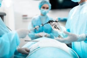
The Baylor study included 187 mesothelioma patients. These patients had PET-CT scans or standard CT scans for mesothelioma staging. But they also had diagnostic laparoscopy. A laparoscope is a camera attached to a thin tube. It lets doctors look inside the chest and abdomen through a small incision.
In 17 percent of the patients, diagnostic laparoscopy revealed that they had mesothelioma in their abdomen as well as their chest. This hardly ever showed up on their imaging scans. PET-CT scans correctly found peritoneal spread only 23 percent of the time. In 77 percent of cases, these patients had negative PET-CT scans for peritoneal disease.
Overall, PET-CT was 68 percent accurate at finding peritoneal disease during mesothelioma staging.
“PET-CT has low sensitivity and diagnostic accuracy to identify peritoneal disease in malignant pleural mesothelioma,” lead author R. Taylor Ripley explains. In an article in the Annals of Thoracic Surgery, Dr. Ripley and colleagues say diagnostic laparoscopy is more likely to find unexpected tumors in the abdomen.
“DL as part of pre-operative staging defines an important subset of patients with bicavitary disease,” Ripley concludes. The report recommends that mesothelioma surgeons make DL a routine part of mesothelioma staging before surgery.
Source:
Ripley, RT, at al, “Diagnostic Laparoscopy Improves Staging of Malignant Pleural Mesothelioma with Routine PET Imaging”, December 2020, Annals of Thoracic Surgery, Online ahead of print, https://www.annalsthoracicsurgery.org/article/S0003-4975(20)32035-X/pdf