MRI Imaging May Be Superior to CT for Pleural Mesothelioma Diagnosis and Staging
 When it comes to diagnosing and monitoring treatment response in people with pleural mesothelioma, MRI imaging may be better than CT.
That is the message from a new Italian study published in the online medical journal Cancers.
RIght now, CT (computed tomography) is the most popular form of imaging for pleural mesothelioma. Cancer doctors use CT images to help diagnose mesothelioma. They also use it to see how widespread the cancer is and to find out if treatment is making a difference.
But radiology researchers at Pisa University Hospital say MRI imaging has advantages over CT for people with the rare asbestos cancer.
Mesothelioma Diagnosis and Prognosis
Malignant pleural mesothelioma is the most serious of several health conditions linked to asbestos. It is most common in people who have lived or worked around this carcinogen. The longer the exposure, the higher the chance that the person will eventually develop mesothelioma.
Most people who get pleural mesothelioma have a poor prognosis. One reason is that mesothelioma is challenging to diagnose. By the time most people get a definitive diagnosis, the cancer may be very advanced.
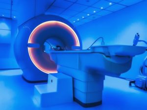
Tools like CT or MRI imaging can help with mesothelioma diagnosis, staging, and treatment planning. They give doctors a non-invasive way to look inside the body. CT is more common than MRI. But MRI imaging provides more information than a CT scan.
The authors of the new study say that additional information lets doctors make better use of advanced computer-based diagnostic, staging and prognostic tools.
CT versus MRI Imaging
CT (computed tomography) imaging uses X-rays. A CT scanner takes a series of X-ray images of a mesothelioma tumor. A computer puts the images together to create a 3-dimensional image of the whole tumor.
Magnetic resonance imaging (MRI imaging) does not use ionizing radiation. It creates images using a powerful magnetic field and radio waves. MRI can produce clearer, more detailed pictures of soft tissues than CT scans do.
That is especially important for mesothelioma tumors which are usually flat and irregularly-shaped. And the extra detail in MRI images makes it possible for a computer to perform even more advanced evaluation.
“Computer-based methods can make feasible tasks like segmentation that would otherwise be impracticable,” says lead author Chiara Romei of Pisa University Hospital. “MRI, thanks to its high soft tissue contrast, evaluation of contrast enhancement, and through diffusion-weighted-images, could replace CT in many clinical settings.”
CT and MRI imaging can assist with mesothelioma diagnosis. But the only way to know for sure if a patient has malignant mesothelioma is for a pathologist to examine some of the tumor cells under a microscope.
Source: Romei, C, et al, “New Updates of the Imaging Role in Diagnosis, Staging, and Response Treatment of Malignant Pleural Mesothelioma”, August 30, 2021, Cancers, https://www.mdpi.com/2072-6694/13/17/4377