Protease Imaging May Track Mesothelioma

A chemical that turns certain extracellular enzymes fluorescent may offer an effective, non-invasive method for tracking the progression of malignant pleural mesothelioma.
Pleural mesothelioma is an aggressive cancer of the membrane around the lungs. Because mesothelioma tumors tend to be thin and spread out, they are more difficult to ‘see’ with standard imaging methods than some solid tumors. Sometimes, surgery is the only reliable way to determine how fast and how large a mesothelioma tumor is growing.
But researchers at Penn State and the University of Pennsylvania, one of the country’s most active mesothelioma research centers, say they have successfully used an optical imaging agent called ProSense 680 to measure the size of mesothelioma tumors in living mice.
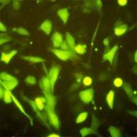
ProSense 680 is a chemical that binds with extracellular cysteine protease enzymes, important cell-signaling enzymes that are present in larger amounts where there are cancer cells, and makes them fluorescent. Theoretically, the more cancer cells are present, the more cysteine protease activity there will be. When ProSense 680 was injected into mice that had mesothelioma cells grafted just under their skin, the cysteine proteases lit up, making it possible for them to be seen and measured with special fluorescent imaging equipment.
To make certain that what they were seeing was, in fact, cysteine protease activity, researchers tested the cysteine protease activity of the cells using other methods, as well. They concluded that, “ProSense 680 yielded a robust tumor signal in malignant pleural mesothelioma subcutaneous grafts… Optical imaging of extracellular cysteine protease activity is useful for tracking malignant pleural mesothelioma tumor cell mass in vivo.” They go on to say that the same method is also feasible for tracking mesothelioma cells that have spread into the abdomen.
Source:


