Ultrasound: A Safer Way to Manage Mesothelioma?
The same technology used by obstetricians to track pregnancies and by cardiologists to find blood clots may also play a valuable role in managing mesothelioma.
An Indonesian mesothelioma doctor says ultrasound technology is not only safer than radiation, but also portable, non-invasive and relatively inexpensive to use. In a recent article published in an Indonesian medical journal, Dr. C.M. Rumende of the University of Indonesia Medical School says ultrasonography of the lungs allows clinicians to diagnose some abnormalities common in mesothelioma, including the buildup of lung fluid known as pleural effusion, more rapidly than they could with other imaging modalities.
“In addition to pleural effusion,” observes Dr. Rumende, “other lung disorders can be diagnosed by ultrasound such as peripheral lung tumors, other pleural abnormalities caused by pleural fibrosis and tumor metastasis, as well as the pleural tumor (mesothelioma).”
Ultrasound imaging can also be used to safely guide surgeons during invasive lung procedures such as aspiration of pleural fluid, tissue biopsies of mesothelioma tumors and chest tube insertion. By allowing surgery to be very precise, ultrasound can increase the likelihood of surgery success and reduce the rate of complications.
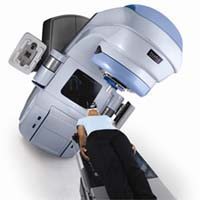
Pleural mesothelioma, an aggressive cancer of the membrane surrounding the lungs, is often difficult to diagnose. Many of the methods commonly used to find and track it, such as MRI/PET or CT, involve the use of internal radioactive tracers or external radiation. In contrast, ultrasound imaging uses only sound waves so mesothelioma patients are not exposed to radiation.
Advances in technology have allowed ultrasound machines to become much smaller (including hand-held units) and more portable. At a fraction of the cost of radiation-based technologies, these smaller ultrasound machines may be an increasingly important mesothelioma management tool, particularly in poorer countries where rates of the disease are continues to rise.
However, ultrasound does have limitations. According to Dr. Rumende, the technology is not very good at detecting tumors and other abnormalities in the mediastinum, the central compartment of the thoracic cavity.
Sources:
Popic, Ramac, “The possibilities and limitations of dirct digital radiography, ultrasound and computer tomography in diagnosing pleural mesothelioma”, December 2012, Collegium Antropoligicum, pp. 1263-1271.