Promising Technology for Mesothelioma Diagnosis, Prognosis and Treatment Planning

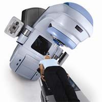
Patients diagnosed with or suspected of having malignant pleural mesothelioma would do well to seek treatment in a center with access to a hybrid nuclear imaging modality called PET/CT.
That’s the conclusion of a group of medical researchers in India who studied the value of this new integrated imaging technique for mesothelioma and recently published their findings in the journal Molecular Imaging and Biology.
Positron Emission Tomography (PET) is a nuclear imaging technique which tracks a positron-emitting ‘tracer’ in the body in order to produce three dimensional images of functional processes. The most common biologically active molecule used for this scan is Fludeoxyglucose (FDG), which is an analogue of glucose. Images are produced as energy is given off during metabolism of the FDG.
Computed Tomography, or CT scanning, uses X-rays to produce images of internal structures. A series of photos or ‘slices’ are combined to create a 3D effect. Combining PET and CT technologies into an integrated system can give doctors more information about a patient’s mesothelioma than either technology can do by itself.
Mesothelioma is an aggressive cancer of the mesothelium, a membrane that forms the lining of several body cavities. In an analysis of the medical literature, the Indian team found that PET/CT showed promise in five crucial areas of mesothelioma management: 1) differentiating malignant pleural mesothelioma from benign lung diseases, 2) staging the cancer in order to determine a patient’s suitability for surgery, 3) evaluating how well the cancer is responding to a given treatment, 4) assessing the patient’s prognosis based on how tumor cells metabolize the FDG, and 5) planning for radiotherapy. Of these, cancer staging and ongoing monitoring of tumor progression are the most common uses for PET/CT.
“In all of these areas, PET/CT convincingly shows better results than conventional anatomic imaging alone and thereby can aid in exploring novel therapeutic approaches,” report the study’s authors.
Differentiating malignant pleural mesothelioma, predicting prognosis, and planning for radiotherapy are less common uses for PET/CT technology, but the researchers say preliminary evidence suggests they are worth further study. In addition to examining the value of PET/CT the article also takes a closer look at using PET imaging alone and an advanced algorithm to define the borders of the mesothelioma, determine its volume and assess how metabolically active it is.
They write, “This global disease assessment, we believe, will be the way forward for assessing this malignancy with a non-invasive imaging modality.”
Sources:
Basu S, Saboury B, Torigian DA, Alavi A “Current Evidence Base of FDG-PET/CT Imaging in the Clinical Management of Malignant Pleural Mesothelioma: Emerging Significance of Image Segmentation and Global Disease Assessment”, Dec 7 2010, Molecular Imaging and Biology. Epub ahead of print.
Ambrosini V et al, “Additional value of hybrid PET/CT fusion imaging vs. conventional CT scan alone in the staging and management of patients with malignant pleural mesothelioma.” August, 2005. Nuclear Medicine Review, Central and Eastern Europe.


