CT Discovery May Lead to Earlier Mesothelioma Diagnosis
Researchers in Western Australia believe they may have found a way to overcome one of the biggest challenges in the management of malignant pleural mesothelioma—making the diagnosis earlier.
Malignant mesothelioma is a fast-growing cancer that starts on the membrane, or pleura, that surrounds the lungs.
Because the seeds for this disease can exist for decades before symptoms develop, and because the early symptoms of mesothelioma tend to be vague and similar to those of other conditions, the so-called “asbestos cancer” often goes undetected until it is in an advanced stage. As a result, the survivability of this disease is typically very low.
But scientists at the University of Western Australia in Perth say there may be a simple way to enhance the accuracy of CT scanning for pleural mesothelioma and detect the disease earlier. The key is to fill the chest cavity with air prior to the test.
Mesothelioma Diagnosis: CT Has Limits
Pleural mesothelioma is an aggressive cancer that requires quick action on the part of doctors and patients to stop it from spreading.
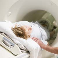
Even though the disease is highly resistant to most cancer treatments, early diagnosis can improve the odds of survival. In conjunction with blood tests, exams, and work histories, CT scanning is a critical component of pleural mesothelioma diagnosis.
But, as the Australian research team observed, CT often fails to detect smaller nodules on the pleura. They believe this may be because there is not enough difference between the density of these nodules and the density of the pleural fluid that surrounds them.
On CT scan, these nodules, some of which could be early mesothelioma tumors, may simply be “lost” in the pleural fluid.
Air Could Improve Mesothelioma Diagnosis
But there may be a work-around. In a recent article in the journal Chest, doctors detail the cases of six patients with pleural abnormalities that were virtually undetectable on CT until their chests were filled with air (known as pneumothorax)—either deliberately or by accident because of a leak.
“Pneumothorax after pleural fluid drainage permitted the visualization of small pleural abnormalities on CT scan, which would be amenable to image-guided biopsies,” writes lead researcher Edward Fysh, PhD.
Dr. Fysh and his colleagues say the cases provide “proof of principle” that the sensitivity of CT to detect pleural abnormalities like mesothelioma tumors depends on the density of the surrounding tissue and could be enhanced by intrapleural air.
They conclude their article by calling for formal study of the technique as a method for improving the diagnosis of mesothelioma and other pleural diseases.
Sources:
Fysh, ETH, et al, “Air in the Pleural Cavity Enhances Detection of Pleural Abnormalities by CT Scan”, June 2018, Chest, pp. e123-e128