Mesothelioma-Related Lung Fluid: Better Diagnosis with HIgh Tech Tool
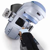
 Technology that integrates 18-FDG PET and CT imaging together may be a more powerful way to diagnose malignant lung fluid in people with pleural mesothelioma than either modality alone.
Scientists in China did a retrospective study to compare the imaging results of people with pleural effusion from various causes, including malignant pleural mesothelioma.
The team found that the integrated PET/CT technology accurately diagnosed malignant pleural effusion better than 18FDG-PET imaging alone and much better than CT imaging.
What is Malignant Pleural Effusion?
Malignant pleural effusion is a condition where fluid containing cancer cells collects in the pleural space between the lungs and the chest wall. If this fluid is caused by a condition other than malignant mesothelioma or another cancer, it can be benign, meaning it does not contain cancer cells.
Recognizing whether this fluid is malignant or benign is an important part of pinpointing the underlying condition that is causing it and making plans for treatment. As many as 90 percent of pleural mesothelioma patients experience malignant pleural effusion, which can cause chest pain, shortness of breath, and fatigue.
Distinguishing Benign from Malignant Effusion
Identifying when pleural effusion is malignant can be an important step toward making a diagnosis of malignant mesothelioma or another type of cancer. Several cancers, including ovarian, breast, and lung cancer, tend to cause pleural effusion when they metastasize or spread.
The new study conducted at Harbin Medical University found that integrated 18-FDG PET/CT accurately detected malignant pleural effusion in 93.5% of cases, while CT studies alone had a sensitivity of just 75%. 18-FDG PET alone returned an accurate positive result in 91.7% of cases.
“18-FDG PET/CT integrated imaging is a more reliable modality in distinguishing malignant from benign pleural effusion than 18-FDG PET imaging and CT imaging alone,” writes lead author Yajuan Sun in the online open journal PLoS One.
Mesothelioma patients and others with malignant pleural effusion were found to have nodules or irregular pleural thickening on CT imaging and a higher uptake of the 18-FDG tracer on PET imaging.
Because malignant mesothelioma is so rare, the technology and expertise needed to accurately identify and treat it is not always readily accessible. For the best mesothelioma outcomes, mesothelioma patients and their families are urged to look for care centers that have the most experience dealing with asbestos cancer.
Source:
Sun, Y, et al, “The Role of 18-F-FDG PET/CT Integrated Imaging in Distinguishing Malignant from Benign Pleural Effusion”, August 25, 2016, PLoS One


